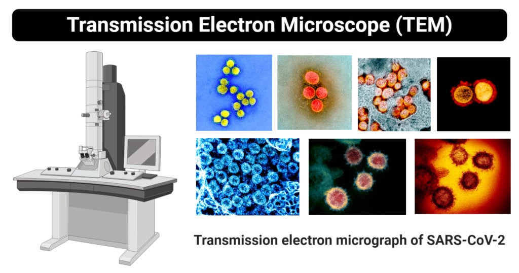

In the STEM method developed by the researchers, the sample stands still while the microscope sends two beams of electrons tilted against each other, and two detectors are simultaneously used to record the signal. Finally, taking multiple images with TEM requires shooting a beam of electrons though the sample each time, and the total dose can actually affect the sample's structure during acquisition and produce a false or corrupted image. The 3D images generated in this way are also prone to artefacts, which are difficult to remove afterwards. The problem with this approach is that it requires extreme precision on hundreds of images, which is hard to achieve. The images can then be reconstructed on a computer to gain a 3D representation of the sample. A way around this problem is to acquire consecutive images while rotating the specimen through a tilt arc. However TEM only provides 2D images, which are not enough for identifying the 3D morphology of the sample, which often limits research. TEM is a very powerful technique that can provide high-resolution views of objects just a few nanometers across - for example, a virus, or a crystal defect. For example, it can be curvilinear, like DNA or the mysterious defects that we call 'dislocations', which govern the optoelectronic or mechanical properties of materials." "But in some cases with TEM we know something about what shape the sample's structure must have. "Our own eyes can see 3D representations of an object by combining two different perspectives of it, but the brain still has to complement the visual information with its previous knowledge of the shape of certain objects," says Hébert. Furthermore, it can rapidly provide a "sense" of three dimensions, just like we would have with a 3D cinema. The novelty of the method is that it can acquire images in a single shot, which opens the way to study samples dynamically as they change over time. The technique, developed by EPFL researcher Emad Oveisi, relies on a variation of TEM called scanning TEM (STEM), where a focused beam of electrons scans across the sample. The labs of Cécile Hébert and Pascal Fua at EPFL have developed an electron microscopy method that can obtain 3D images of complex curvilinear structures without needing to tilt the sample.


 0 kommentar(er)
